Get Exposure Before You Start Pulling
Reduce stomach gently, divide short gastrics when needed, expose both crura, and identify the sac. Pulling on the esophagus before the sac is free is how you create a problem.
Type I-IV hiatal hernias, paraesophageal hernia strategy, mediastinal dissection, esophageal length, cruroplasty, mesh reinforcement, recurrence, volvulus, gastropexy, documentation, CPT, and wRVUs.
Type I-IV hiatal hernias, paraesophageal hernia strategy, mediastinal dissection, esophageal length, cruroplasty, mesh reinforcement, recurrence, volvulus, gastropexy, documentation, CPT, and wRVUs.
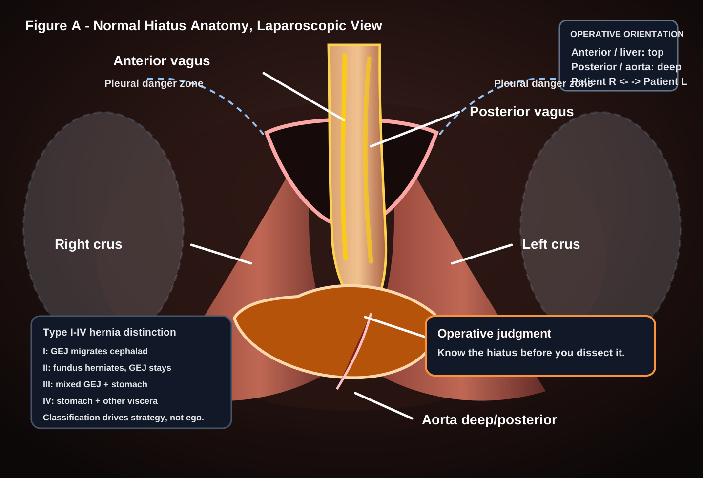


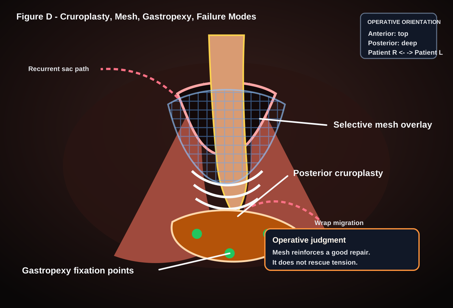
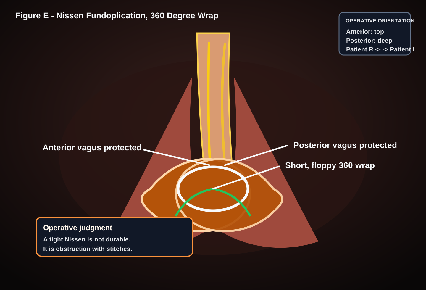
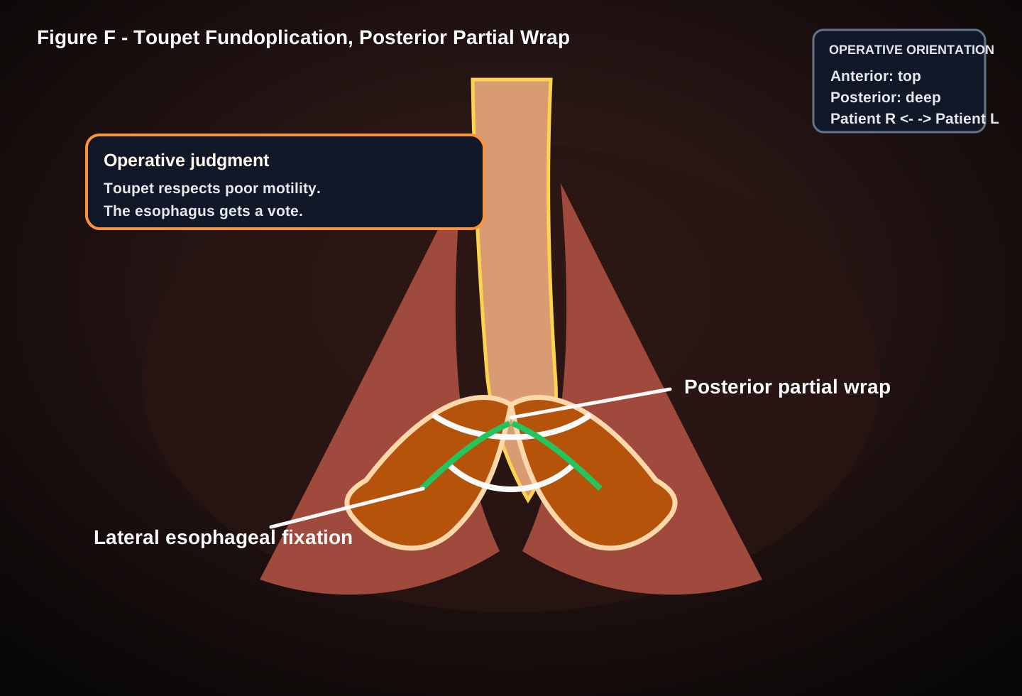
Real operative video integration is intentionally curated, not scraped. Until an approved public or Graydon-owned video is embedded, use the atlas above as the key-frame map and review any operative footage against these moments: sac reduction, circumferential mediastinal mobilization, length assessment, crural closure, selective mesh, gastropexy, and wrap geometry.
Right crus, left crus, anterior vagus, posterior vagus, phrenoesophageal membrane, left gastric pedicle, short gastrics, pleura, pericardium, aorta, and the GE junction.
Type I is sliding GE junction migration. Type II is true paraesophageal herniation with GE junction in place. Type III combines both. Type IV brings stomach plus another viscus into the chest.
The safe plane is on the hernia sac, not in the esophageal wall. Circumferential mobilization should create a posterior window and restore at least 2.5-3 cm of tension-free intraabdominal esophagus.
Recurrence usually reflects missed sac, inadequate length, crural tension, poor tissue, wrap migration, or failure to anchor the stomach when gastropexy was the better operation.
Postprandial pain, early satiety, regurgitation, dysphagia, dyspnea, aspiration, anemia from Cameron lesions, or impaired quality of life.
Gastric volvulus, obstruction, ischemia concern, bleeding, or inability to decompress. The first decision is source control and viability, not perfect elective reconstruction.
GERD with objective reflux, anatomy suitable for repair, and a workup that supports an antireflux operation rather than isolated medical therapy.
Recurrent hernia, slipped wrap, obstructing wrap, recurrent volvulus, or mesh/wrap complication after careful imaging, endoscopy, and physiology review.
Reduce stomach gently, divide short gastrics when needed, expose both crura, and identify the sac. Pulling on the esophagus before the sac is free is how you create a problem.
Perform complete circumferential mediastinal mobilization, protect both vagus nerves, stay on sac, and develop enough length that the GE junction sits below the crura without traction.
If you cannot get 2.5-3 cm of tension-free intraabdominal esophagus after real mobilization, call it a short esophagus. A tight wrap on a short esophagus is a recurrence plan.
Posterior cruroplasty is standard. Calibrate around the esophagus, assess tissue quality, and avoid making dysphagia the price of a pretty hiatus.
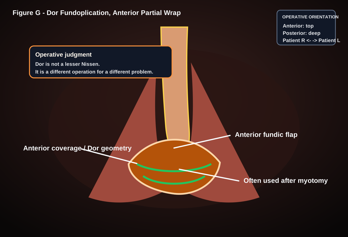
Nissen requires acceptable motility and a short floppy wrap. Toupet is safer for weak motility or dysphagia risk. Dor is a different tool, often used when anterior coverage is the goal.
Mesh is for selected large defects, poor crura, or recurrence risk. Gastropexy is for volvulus control, frailty, high-risk recurrence, or when a full repair is unsafe.
Leaving sac behind and calling the stomach reduced.
Failing to recognize a true short esophagus.
Building a wrap under tension.
Injuring pleura and ignoring capnothorax physiology.
Overtight cruroplasty causing postoperative dysphagia.
Using mesh as a substitute for mobilization.
Tell anesthesia, reduce insufflation pressure, watch ventilation and hemodynamics, decompress if tension develops, and place a drain/chest tube only when physiology or injury requires it.
Mobilize more before declaring failure. If length remains inadequate, consider Collis gastroplasty or referral/staging rather than forcing a high-tension repair.
Identify prior wrap and mesh, use EGD liberally, preserve the vagus nerves if possible, and convert before esophageal injury becomes the operation.
Reduce, assess viability, decompress, and consider anterior gastropexy or PEG gastropexy when definitive reconstruction is more dangerous than recurrence.
Recognize intraoperatively, repair in healthy tissue, test with EGD or air leak, drain, consider stent when appropriate, and do not hide the injury in the op note.
Separate edema from mechanical obstruction. Start with diet modification and imaging/endoscopy; dilate selectively and revise only when anatomy proves the repair is the problem.
Consider tight wrap, vagal injury, or unmasked gastroparesis. Manage diet and symptoms first, then define anatomy before contemplating revision.
Work up mechanism with CT/esophagram/EGD. Recurrence from tension, missed short esophagus, or failed crura needs a different plan than simple recurrent reflux.
Rare but serious. Define location endoscopically, involve experienced foregut help early, and plan reoperation around esophageal preservation rather than mesh removal alone.
Usually from short gastric or fundus traction. Pack, expose, control the source, and convert if visualization is not good enough to make the next move safely.
Click any card to reveal the answer. Use filters for student, resident, and advanced decision-making review or Quiz Mode for one-question-at-a-time board prep.
Six realistic case stems that teach operative judgment, prioritization, bailout thinking, and documentation discipline.
These cases are educational examples for surgical learning and documentation awareness. They are not patient-specific medical advice. Actual management depends on patient physiology, anatomy, local resources, attending judgment, and institutional protocols.
Values below are pulled from the FreeCPTCodeFinder CPT database at runtime so RVUs and estimated Medicare payments stay aligned with the coding engine.